Search Our Website
80 results found with an empty search
- 2025 Update on PPID (Cushing’s Disease)
Pituitary pars intermedia dysfunction (PPID) — also known as equine Cushing's disease — is an age-related, slowly progressive, degenerative disease that affects many horses. Typical signs that your horse may have PPID include a long, wavy hair coat that doesn’t shed, excessive thirst and urination, muscle wasting, and increased susceptibility to infections and laminitis. If you suspect that your horse may have pre-Cushing’s or Cushing’s disease, it’s important to discuss this with your veterinarian and begin a treatment plan that may include medication as well as dietary management and general wellness care to promote longevity and ensure a good quality of life. The Equine Endocrinology Group (EEG) is composed of experts who provide advice in the form of written guidelines to help veterinary practitioners diagnose and manage equine endocrine disorders. Guidelines are updated every two years. Here are the most recent guidelines, released in October 2025: Learn more: https://www.bicanadaequine.ca/ppid/ppid-symptoms
- Cresty Necks and Laminitis: Equine Endocrinology
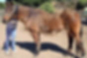
By Nora Grenager, VMD Updated in 2020 (originally published in Bay Area Equestrian Network September 2006) Example of severe chronic laminitis The two main endocrinologic concerns in horses are equine Cushing’s disease (also known as pituitary pars intermedia dysfunction) and equine metabolic syndrome (also known as “insulin dysregulation” or, formerly, “insulin resistance”). These disorders are increasing in prevalence as our equine population is better cared for and living longer, and improved diagnostics are available. There are two main similarities between these conditions: 1) insulin dysregulation, and 2) the potentially devastating possibility of laminitis. While it is an area of much research, there are numerous theories as to why these horses are predisposed to laminitis, and it is beyond the scope of this article to discuss them all. However, laminitis is likely related to insulin dysregulation, which is a reduced ability of the body to respond appropriately to insulin released by the pancreas after eating a meal. It is important to understand the signs of insulin dysregulation and be able to effectively manage these horses to decrease the risk of laminitis. Equine Cushing’s Disease Very hirsute horse The cause of equine Cushing’s disease is a benign tumor, likely caused by oxidative stress, in a part of the horse’s brain called the pituitary gland. It is not a typical neoplastic tumor, rather a lack of regulation of hormone secretion due to alterations in local factors in that region of the brain. This alteration (namely, a decrease in local dopamine production) leads to unregulated hormone secretion by the pituitary gland, enlarging this area such that it forms a tumor. This tumor secretes a variety of hormones, including one called adrenocorticotropic hormone (ACTH) that causes the adrenal gland (in the abdomen) to release increased amounts of steroid (cortisol). Other secreted hormones include α-MSH, POMC, and β-endorphin. The increased levels of these circulating hormones cause the typical clinical signs , including: a long, wavy hair coat that does not completely shed out; abnormal fat distribution, such as a cresty neck or fat pads above the tailhead; weight loss, especially over the ribs; muscle loss, along the top line or leading to a pot belly; recurrent laminitis; lethargy and exercise intolerance; increased sweating, increased water intake and urination; and immune system suppression or recurrent infections. Many horses with Cushing’s disease also have chronic insulin dysregulation. The most common clinical signs are the abnormal, long hair coat (called “hirsutism”) and chronic laminitis. However, it is important to remember that horses with milder disease can have milder signs, and a long haircoat may not be seen in all horses with Cushing’s. Equine Cushing’s disease is more common in older horses (in their late teens to 20s), but can be seen in horses as young as 7 years old. There are some breeds that seem to be more predisposed, and ponies also seem to be predisposed to the condition. Example of a cresty neck Definitive diagnosis of equine Cushing’s disease can be challenging. It is relatively easy to diagnose in an older horse with more severe disease and typical clinical signs, such as a long hair coat and laminitis. In these cases, in which the horse has the typical clinical signs of equine Cushing’s disease, diagnostic tests are sometimes skipped because the signs are so suggestive. Remember, though, that having lab values to monitor is very helpful so testing should be done when possible. Yet sometimes a “diagnostic therapeutic” challenge is used, in which we try treating a horse for 30-60 days and monitor the response; the treatment for Cushing’s (pergolide) is so effective and safe that we have our answer if the horse improves on medication. There are several tests available. The difficulty with all the available tests is that they can be negative in horses with early Cushing’s disease; thus, if the index of suspicion is very high, we may still opt to treat the horse even if the test is negative. The first recommended test is measurement of the resting levels of ACTH, one of the hormones that is overproduced by the tumor in the pituitary. This test requires one blood sample. While it is fairly accurate, it still can have false negatives (i.e., about 30% of the time horse with Cushing’s disease has a negative test result). If the resting ACTH levels are normal, the next recommended test is called the thyrotropin releasing hormone stimulation test (the TRH stim test). This test is slightly more sensitive (i.e., more likely to correctly diagnose the horse with Cushing’s), but requires two blood samples, taken 10 minutes apart, and intravenous administration of TRH. The test is very safe but is obviously a bit more expensive than just measuring the resting ACTH once. The dexamethasone suppression test is another test that is slowly falling out of favor as the ACTH testing has been shown to be more reliable. This test requires two veterinary visits: at the first visit, blood is drawn to measure the cortisol level and a dose of dexamethasone is given. At the second visit, about a day later, blood is drawn to again measure the cortisol level. The test relies on a normal endocrine negative feedback loop. In a normal horse, a dose of dexamethasone (steroid) suppresses the pituitary gland, telling the gland that it is unnecessary to make more cortisol (steroid made by the body) because some has just been given. Therefore the second sampled blood level of cortisol is very low in a normal horse. In a horse with Cushing’s, the tumor in the pituitary gland results in production of cortisol no matter what levels of steroid are in the body, so it does not respond to the dexamethasone the veterinarian administers; therefore the second level of cortisol is not significantly different from the first, pre-dexamethasone, level. This test can also have false positive or false negative results. There are two potential drawbacks: first, it necessitates two visits from your veterinarian, which increases cost. Second, it involves the administration of steroid to a horse that is already possibly predisposed to laminitis. Therefore there is a very low risk of precipitating a bout of laminitis. Most practitioners feel, however, that the benefits of knowing the test results often outweigh the risks. It has recently been shown that the tests for equine Cushing’s disease are even more sensitive in the late summer/early fall (i.e., mid-August through mid-October); therefore, veterinarians may recommend testing at that time. A variety of other tests exist, and there is ongoing research to develop even more sensitive and specific ways to diagnose equine Cushing’s disease in those horses in which the diagnosis is difficult. The astute reader may be asking why we do not just measure cortisol levels, since the overproduction of that hormone is a hallmark of the disease. Unfortunately, the daily variations in cortisol levels render this measurement useless. Evaluation for insulin dysregulation should always be performed with with tests for Cushing’s disease since many horses with Cushing’s have concurrent insulin dysregulation. Discussion on the diagnosis of insulin dysregulation is in the following section. Treatment is advocated because of the possibly devastating effects of laminitis and immune suppression. Fortunately, there is a highly effective specific treatment for equine Cushing’s disease called “pergolide.” Pergolide acts like dopamine and works on the pituitary gland to prevent the release of excess hormones from the tumor. It is given as a tablet once daily for the rest of the horse’s life. Most of the time a beneficial response is seen in 4–6 weeks, but sometimes the dose needs to be increased or decreased. Pergolide has virtually no known side effects in horses at appropriate dosing levels; some horses will become mildly inappetant when the medication is first started. The appetite returns in these horses when they are started on a lower dose and it is gradually increased. Often bloodwork is repeated to help monitor response to treatment, and generally the response to treatment is easily seen. The trade name for pergolide is “Prascend®”—while there are compounded versions of pergolide that are less expensive, they are considerably less reliable and effective, so in the long run end up costing more for less effect. Several other drugs (such as cyproheptadine and trilostane) have been used to treat equine Cushing’s disease, but none have been shown to have the efficacy of pergolide. Occasionally one of those drugs is added to pergolide treatment in severe cases that need additional therapeutic help. Many nutritional or herbal supplements are also available (often including magnesium and chromium or chasteberry), and may have variable efficacy; however, none have been proven to be safe and effective. Often these horses also have insulin dysfunction, so treatment for that will be similar to what is described in the next section. The proper treatment of Cushing’s disease with pergolide eventually alleviates many of the signs of insulin dysregulation since the underlying cause is being treated. In addition to daily medication, some feed and management changes should be considered. These horses need regular farrier care, good dental care (immune suppression and older age make them prone to dental problems and secondary sinus infections), routine preventive veterinary care including fecal egg counts, and good quality feed. Some horses may need to be clipped to help prevent hyperthermia. High starch/sugar feeds should be avoided in the horses that have insulin dysregulation to help minimize fluctuations in glucose levels. More about nutrition for horses with insulin dysregulation is written in the following section on metabolic syndrome. Horses with Cushing’s that do not have documented insulin dysregulation but suffer from weight loss will have different dietary needs. Equine Metabolic Syndrome Equine Metabolic Syndrome is defined as chronic insulin dysregulation, obesity or abnormal fat distribution (e.g., a cresty neck; fat pads over the tailhead, eyes, and withers), and increased risk of laminitis in horses that do not have equine Cushing’s disease. This is a relatively newly recognized condition, and there is a lot of ongoing research about this disorder and our understanding of it continues to grow. Horses with this condition tend to be younger than horses with Cushing’s disease. These horses have documented insulin dysregulation and sometimes have increased circulating fat levels. Example of horse with metabolic syndrome. Note the cresty neck and excess fat by the tailhead. The cause of metabolic syndrome is not fully understood, but several theories are being closely considered. First, obesity leads to insulin dysregulation (much like people with Type II diabetes). Second, insulin dysregulation develops secondary to abnormal nutritional usage in genetically predisposed animals. Third, fat cells can be metabolically and hormonally active and alter insulin usage in a peripheral Cushinoid-type syndrome. It is logical to think that horses with metabolic syndrome will perhaps transition into equine Cushing’s disease as they age; however, this has not been proven. There are several ways to test for insulin dysregulation associated with metabolic syndrome (and for horses with Cushing’s disease). The most basic, first-line, test is to measure resting fasting glucose and insulin concentrations first thing in the morning before breakfast. Basically, a normal horse should have low insulin levels when glucose levels are normal. A horse with insulin dysregulation has increased levels of insulin (to try to make up for tissue resistance to it). The second, slightly more sensitive, test for insulin dysregulation involves administering a specific amount of light Karo syrup, then testing the blood a specific amount of time later (approximately 1 hour) to monitor the response. A third, more involved, test is the combined glucose-insulin test (CGIT), which is used when insulin dysregulation is suspected but the first two test results are normal. The CGIT involves placing an intravenous catheter, administering glucose and insulin, and measuring the body’s response to this at multiple time points. There is no one specific treatment available for equine metabolic syndrome regardless of whether it is definitively diagnosed or not. Treatment focuses on the management of the insulin dysregulation, including reducing the horse’s weight, dietary management, and increasing exercise. The most important goal of dietary management is to restrict the amount of soluble carbohydrates (often called “nonstructural carbohydrates”) in the feed, because they alter the insulin levels the most. Grazing should be discussed with your veterinarian and many owners use grazing muzzles or dry lots; however, it is not recommended to lock the horse in a stall. Hay can be analyzed (your vet can direct you to a company that will do this) for its soluble carbohydrate content; it is worth doing this if you purchase large amounts of hay at a time. Some types of grass hay have fairly low soluble carbohydrates, and should be fed at 1.5 to 2% of body weight, as directed by your veterinarian. Oat hay should be avoided. If the horse’s hay intake is tapered down to 1% of its body weight and it is still having difficulty losing weight, soaking the hay is a way to further decrease the soluble carbohydrates. It should be soaked in warm water for 20 minutes, or cold water for 60 minutes, immediately prior to feeding. A vitamin/mineral supplement should be fed, and if additional calories are needed they should be provided by sources high in fat and protein, rather than carbohydrates, such as corn oil or rice bran. Sweet feeds, apples, carrots, and high sugar treats should be minimized or, more often, completely removed from the diet. Most feed companies also make low-carbohydrate feeds specifically designed for horses with insulin dysregulation. Unless a horse is currently dealing with a bout of laminitis, increasing exercise is essential to reducing body fat. Some studies have shown that increased exercise also might help improve tissue insulin sensitivity. If a horse is on a good exercise program with a proper diet and still not losing weight, or is in a current bout of laminitis, thyroid hormone supplementation may be recommended by your veterinarian to improve insulin sensitivity. Many horses with insulin dysregulation used to be incorrectly categorized as “hypothyroid.” Further research has shown that very few horses are actually hypothyroid, however many horses with insulin resistance can have low measurable levels of thyroid hormone. However, their thyroid glands are working fine, and the insulin resistance is the main issue. In spite of this, supplementing with thyroid hormone can help boost the metabolism, improve insulin sensitivity, and get a horse to start losing weight. The thyroid hormone is typically given once daily for a couple of months and then tapered down and discontinued. A drug used to treat insulin resistance in humans, called “metformin,” is also used in horses with severe insulin dysregulation. It is typically given 30 minutes prior to feeding and can only be used for a few weeks at a time. Many other medications taken from the human medical field have also been evaluated and tried though many are cost prohibitive in the majority of cases at this time. Also, just as with equine Cushing’s disease, there are many herbal supplements advocated to treat equine metabolic syndrome. Again, there is little proof of efficacy or safety of these treatments, though anecdotally there are some that are beneficial. If you are concerned that your horse has signs of either metabolic syndrome or equine Cushing’s disease, set up an appointment with your veterinarian for an examination and discussion. It is not always easy to distinguish between these two conditions, and they can often be concurrent. Early recognition is critical to provide your horse with the best care and prevent the potentially devastating disease of laminitis. Ideally, endocrine testing would be part of the annual physical exam in all horses.
- Professional Horse Haulers Based in Central and Northern California
Whether you need to transport your horse across the county or across the country — to a new home, to a show, or for medical care — it’s good to know that there are several professional horse haulers and equine transport experts you can call upon who are headquartered in the Steinbeck Peninsula Equine Clinics service areas in Northern California and near the Central Coast. The list below includes experienced horse haulers who have reported that they carry liability insurance. Photo: Light Star Horse Transportation This is not a comprehensive list. If we missed you and you would like your professional horse transport business included on this list, please email our web team and provide your phone number so we can contact you for details. Please note that Steinbeck Peninsula Equine Clinics is not affiliated with, does not endorse, nor has any business relationships with the individuals, companies, and groups on this list. Also note that descriptions of listed haulers’ services and equipment may not be complete or up to date so please inquire for specifics when you contact them. Light Star Horse Transportation (Terry Konkle) — (877) 254-5112 Hubs in Gilroy, CA and Lexington, KY; specializes in long-distance hauling covering U.S. and Canada Equipment: Ranges from multiple semis for long distances to goosenecks for local/regional trips to smaller pickup trucks with trailers for local deliveries or emergencies Stacie McGrady — (831 )455-5098 Based in Hollister, primarily goes to South Santa Clara County, San Benito County, Monterey County but will go as far north as SF Peninsula (but prefers not to go to East Bay or further north) May be available for night hauling for local emergencies; offers emergency boarding and layovers; worked at SPCA Equipment: Multiple 4-wheel drive trucks and trailers including 3-horse gooseneck and 4-horse bumper pull — both slant load; 2 livestock trailers Alisha Robinson Horse Transport — (707) 292-5153 Based in Cotati; primarily does long hauls in CA, OR, WA and works with JetPets (international equine transport services) Primarily does horse show hauling; does not do short or emergency hauling to clinics Equipment: Head-to-head 7-horse makes 3 boxes / 2 more 7-horses that are reverse slants and make 4 boxes / 8-horse / 6-horse Turchet Transport ( Giselle Turchet ) — (650) 303-9940 Hubs in Woodside, CA; Los Angeles; Medford, OR; covers all of U.S. and Canada Equipment: 2 semis, 3 4-horse head-to-heads, 8-horse head-to-head — all with ramps; plus 2-horse (or single horse box stall) for emergencies or local deliveries — step-up (no ramp) West Coast Vista, LLC (Mark Vanover) — (831) 234-5930 Based in Watsonville; multiple drivers cover all of California, Oregon, Nevada, Idaho Can call any time; contracts with Santa Cruz SPCA; will do trips to UC Davis to do necropsies Equipment: 6 various sized slant-loads with ramps Ray Woods — (831) 245-5050 Based in Hollister, goes as far east as Sacramento and south to LA Would prefer a call ahead of time, but will take emergencies when able to, and offers night services Equipment: 6-horse Sooner / 4-horse Exiss / 4-horse Hart / 18' livestock / 2-horse slant There are several Facebook Groups (discussion groups) that can also serve as resources regarding horse transport — just a few are listed below, and you can search Facebook for others. These are private groups that you will need to join to read or post reviews, learn about available transport opportunities or post your own needs, and check out other posts. Note that these groups offer services from both professionals and non-professional resources so please do your own careful research: Central and Northern California Horse Transport West Coast Equine Hauling Horse Transport Connect - Hauling, Shipping, Transportation Horse Transport (Verified Animal Transport Businesses) Equine Transportation
- The Itchy Horse: Insect Bite Hypersensitivity
By Zoe Davidson , DVM, MRCVS In California, most any time of year can bring a number of health issues caused by insects to our equine friends. One of the most common conditions is Insect Bite Hypersensitivity. Insect Bite Hypersensitivity (IBH, Queensland itch, sweet itch, equine summer eczema) is the most common pruritic ( itchy) disease of horses. Definitely a common topic of discussion within the equine world, but yet not fully understood and frequently mismanaged. The goal of this article is to provide a quick simple run down of the cause, classic symptoms, and what we can do as vets and owners to keep our horses happy and healthy! Cause Allergic sensitivity to the saliva of Culicoides spp., biting midges; and a variety of insect species. There is a genetic component that does predispose some breeds to a more severe reaction than others. Your horse can develop hypersensitivity and associated symptoms even if you do not see your horse being actively bitten. There is much more than meets the eye going on below the skin surface. Simply put, the severity of the clinical signs is not always correlated with the number and frequency of insect bites, but rather the severity of their individual immune mediated reaction. Certain horses develop a much stronger reaction to the saliva than others, and once their system is primed, worse the clinical signs are. Classic Itchy Horse Symptoms IBH can look different in each horse. Most commonly the symptoms include urticaria “hives”, generalized pruritis (“itchiness”) leading to lesions associated with self trauma (e.g skin thickening, lesions, scaling, crusting, hives, and hair loss, which can result in extreme discomfort.) In severe cases the secondary lesions induced by self -mutilation cause open sores and become infected. Lesions in horses are usually found on the trunk, face, mane, tail, and ears, with locations varying depending on the biting characteristics of the particular insect(s). The external signs can be accompanied by a dull, depressed, lethargic temperament. Luckily, it is one of the most well understood of the allergic skin diseases of the horse which means we have developed very effective management practices to help keep our horses happy and healthy this season! TOP MANAGEMENT TIPS: 1. Catch Early - prevention is always the best cure! Monitor your horse twice daily for the signs mentioned above! 2. Fly Control - prevent the flies from biting! Effective fly spray applied 2X/day Routine sheath cleanings Comprehensive fly mask and fly sheet Clean out stalls 2X/day; place manure piles far from barn Place a fan on your horse's stall door! Fun Fact: Flies are actually pretty weak fliers! 3. Supplements: Platinum Performance Skin and Allergy as well as Kinetic Vet Equishield (Skin and Allergy) have been proven to be effective! Omega-3 fatty acids can aid in reducing skin inflammation. When to call a Vet? Hives accompanied by agitation, elevated respiratory rate, effort and/ or depressed lethargic demeanor, Lesions on the head, neck, mane, tail and or belly (ventral midline) that are being rubbed raw, swollen, runny eyes cough. Sometimes your horse may be reacting to a variety of different stimuli, and in this case a thorough examination and further testing is indicated in order to diagnose and effectively treat. Medical Treatments At this time, there are only a few effective treatments once your horse has developed more extensive IBHS. Our options include antihistamines and corticosteroids, both of which are variably effective and do not come without their own potential side effects. In more severe cases, your vet may discuss drawing your horse's blood for an allergy panel. Subsequently, the exact cause of the allergy can be identified, and a specific immunomodulator developed to help reduce their symptoms. A pricey, length and often not always effective option! With this in view, prevention and management are by far the best methods of keeping your horse comfortable, happy and healthy! If you are concerned about your horse, and eager to discuss any of the above information further, please do not hesitate to reach out. We would love to hear from you! Download a PDF of this article... Related resources you may also be interested in: “Equine Allergies” (Video) by Amanda Hedges, DVM, cVA, CVSMT “Equine Allergies” (Article) by Amanda Hedges, DVM, cVA, CVSMT
- Silicosis: A Tragic Irreversible Lung Condition in Horses
By Matt Durham, DVM, DACVSMR Published in Bay Area Equestrian Network February 2007 In the scenic foothills of the Central Coast of California, there lurks a quiet problem that can lead to severe disease in our horses. Commonly known as ‘chalk rock’, this dusty rock form can cause an irreversible lung condition known as silicosis. Silicosis is a well known occupational disease in humans, typically caused by inhaling rock dust created in mining, masonry work, sandblasting, and many other industries where rock is crushed into an easily-inhaled dust. In horses, the disease was first diagnosed in the late 1970s in the Monterey-Carmel area. Before this time, area veterinarians had noted signs of respiratory disease, but had been unsure of its cause. For years, silicosis in horses was believed to be isolated to this small region. But as awareness of the disease increased, cases started to be diagnosed in other parts of the state. What Causes Silicosis? Briefly, silicosis is caused by inhaling silica dust small enough to travel all the way into the smallest airways and air sacs in the lung. Some sources incompletely describe silicosis as a lung disease caused by inhaling quartz dust. As will be discussed in the section titled Silicates, other crystal forms of silica can also cause silicosis. Once the dust particles become lodged in the lung, the body mounts a strong immune reaction. While bacteria or pollen can be broken down and removed from the lung, silicates can not. This causes an ongoing process which can lead to scarring of the lung, and, in people, certain auto-immune disorders. History In humans, silicosis is a fairly well understood but frustrating disease. As an occupational disease, silicosis may have been recognized by ancient Egyptians, and was described well by Agricola in 1556.Even now, there is no cure. Occupational safety measures have dramatically decreased the incidence of the disease, but cases still occur. The most notorious occupational incident occurred in the early 1930s when at least 700 workers died from silicosis. Workers came from all around to tunnel through a mountain in Gauley Bridge West Virginia, eager for any type of work during the Great Depression. The tunnel was to be used to carry water for a new hydroelectric plant. Although it was well understood at the time that silicosis was a risk if dry-drilling was used, the contractors chose this method over the much safer, but slower, wet-drilling technique. Silicates Roadcut exposing siliceous shale of Monterey Formation Silicates are crystalline forms of the element silica. By far the most widespread crystal of silicate is quartz, which is present in most rock types. The form which causes silicosis in horses is cristobalite. Cristobalite causes a more severe reaction in the lung than quartz does. This crystal is present in abundance in the Monterey/Carmel Valley areas. Geologists refer to this particular deposit of rock as the Monterey Formation. Although this rock type is common in this region, it is present from Point Reyes to San Onofre in the coastal ranges, with outcroppings in the San Joaquin valley, and as far south as Baja California. The Monterey Formation developed in shallow seabeds during the Miocene epoch (between 5 and 20 million years ago) from deposits of microorganisms known as diatoms. Diatoms have a non-crystalline form of silica in their structure which, over millions of years, becomes compressed into crystalline forms. Initially, the crystal formed is cristobalite, but with more time, quartz can be formed. Many of the soils of the Monterey Formation have high levels of cristobalite. These soils are often referred to as ‘chalk rock’ locally, as they tend to be dusty and light. (True chalk is actually limestone, which contains no silica.) Geologists refer to some of the various subsets of the Monterey Formation as porcelanite, chert, siliceous mudstone, and siliceous shale. For the rest of this article, the term siliceous soils will be used. Cristobalite can also be formed through volcanic activity. The Sonoma volcanic soils are a likely source of the cristobalite responsible for silicosis in horses from this region. Exposure Areas with siliceous soils are typically in foothill areas. Often, cases are seen not long after recent construction has disrupted the soil, creating a dusty environment. The horses inhale the dust over days to months or even years. Depending on a variety of factors, affected horses may show no signs or could develop severe respiratory compromise. Rate of exposure and individual immune response probably play the biggest roles in terms of severity of signs. Clinical Signs Affected horses will sometimes develop a cough, an elevated respiratory rate (normal resting rate for a horse is typically around 8-16 breaths per minute), flared nostrils at rest, and/or exercise intolerance. It is very important to remember that these signs are typical of respiratory compromise in general, so are not specific for silicosis. Diagnostics Lung x-rays are the simplest test to perform to diagnose silicosis. Abnormal x-rays exhibit classical signs for silicosis in advanced cases. Early or mild cases may have subtle, non-specific lung x-ray findings. Lung x-ray of a horse, showing severe lung fibrosis from silicosis (normal lung is nearly black on x-rays) Samples of fluid and cells from the lungs can be obtained in two ways: a transtracheal wash, or a bronchoalveolar lavage (BAL). Affected horses may exhibit pink crystals within a type of white blood cell called a macrophage. The crystals are silicate crystals, and the macrophages are the cells that attempt to destroy or remove the particles. At Steinbeck Country Equine Clinic, we typically choose the BAL technique for this diagnosis. Ultrasound is occasionally useful in imaging silicosis cases, particularly with advanced cases. Most silicosis changes are deep within the lung, and the ultrasound beam does not penetrate through the air present within the lung, so less advanced cases do not show up well. Treatment In horses, as in humans, there is no cure for silicosis. Treatment involves removing the horse from the silicate dust as well as from other types of dusty or moldy environments. In mild to moderate cases, short courses of steroids and bronchodilators can get a horse through a flare-up. Affected horses may not be able to regulate their temperature, so it is important during hot weather to provide shade, or other means to create a cool environment. Silicate Associated Osteoporosis In horses living on siliceous soils, certain bone deformities and fractures can develop. Horses with silicate associated osteoporosis can develop signs years after moving away from an area with siliceous soils. This disease typically occurs in horses with a longer-term exposure to silicate dust. The bones develop osteoporosis which, as in people, can predispose to fractures. Horses typically develop bowed shoulders, a swayed back, and often a stiff neck secondary to osteoporosis and secondary arthritis in the cervical spine, although early cases have no obvious outward signs. Horse with silicate associated osteoporosis, showing swayed back and bowing of the shoulder blades. Footing material is siliceous shale. [Image courtesy Dr. Coral Armstrong] Body soreness and exercise intolerance are common in horses with silicate associated osteoporosis, and some horses may develop neurologic signs. Fractures are most common in the spine, ribs, pelvis and shoulder blades. Affected horses may or may not have respiratory signs, but will typically have some degree of respiratory involvement. Diagnosis of silicate associated osteoporosis is simple in advanced cases, where outward signs alone are diagnostic. In mildly affected horses, diagnosis is challenging, because vague lameness or stiffness may be the only signs (as can be seen in countless other disease processes). In these cases, nuclear scintigraphy (bone scan) is very sensitive at detecting the disease. Ultrasound of the shoulder blades and x-ray images of the neck can help define the disease as signs progress, but will often be normal in early cases. Treatment for silicate associated osteoporosis with typical anti-inflammatories such as phenylbutazone is often minimally effective. Sometimes steroids will improve the comfort level, but not always. Intravenous Legend® can help with the arthritis pain in the neck that commonly develops, but does not help with bone pain. Tildren® may help decrease bone pain and possibly stop the progression of osteoporosis. Tildren® is in the same family of drugs as Actonel®, Fosamax®, and Boniva®, which are used to treat osteoporosis in people. Recently, zoledronate (Zometa® or Reclast®) has been used experimentally at UC Davis, with promising results. Prevention of Silicosis Prevention of silicosis requires limiting the amount of inhaled silicate dust. Areas of new construction should be avoided. Dry lot situations should be altered as much as is possible. The ideal solution is to plant grass over the area and irrigate throughout the summer months, but this is often very impractical. Incorporating organic material into the soil can help to retain moisture and minimize dustiness. Wood chips or composted manure are fairly effective and inexpensive methods. Topsoil brought in from an unaffected area may also be effective. Future Directions in Silicosis Study Control of the excessive immune response is the primary direction of research in the human field. In horses, the primary areas of study involve silicate associated osteoporosis. We are currently working in conjunction with researchers at UC Davis on studies to determine the exact nature of the bone disease. Currently, Dr. Murray is studying two blood tests, which appear to be promising in detecting the disease process. With earlier detection and monitoring of bone density, we hope to be able to prevent the catastrophic fractures and other bone-related pain.
- AAEP Vaccination Guidelines
The AAEP Vaccination Guidelines are intended to be a reference for veterinarians who utilize vaccines in their respective practices. They are neither regulations nor directives and should not be interpreted as such. It is the responsibility of attending veterinarians, through an appropriate veterinarian-client-patient relationship, to utilize relevant information coupled with product availability to determine optimal health care programs for their patients. Based on the professional judgment of those involved with the development of these guidelines, the recommendations for vaccine administration in this document may differ from the manufacturer’s recommendation. However, it is incumbent on each individual practitioner to reach a decision on vaccine usage based on the circumstances of each unique situation and his or her professional experience. Information provided in these guidelines addresses only those products licensed by the United States Department of Agriculture (USDA) for use in horses (including draft and pony breeds). There are limited data regarding the use of vaccines in other equidae (i.e. asses, donkeys, mules, miniature horses, and zebra); vaccination of these animals is at the discretion of the attending veterinarian.
- Which Vaccinations Should My Horse Get?
The American Association of Equine Practitioners (AAEP) provides guidelines on equine vaccines to assist veterinarians and horse owners in making informed decisions regarding their horses' health. These recommendations are based on the risk of disease, prevalence, geographic location, and the horse's individual circumstances. Here are some commonly recommended vaccines and their rationale: Core Vaccines: The AAEP recommends core vaccines for all horses regardless of their geographical location or exposure risk. These vaccines protect against diseases that pose significant health risks and have a high potential for transmission. Core vaccines include: Eastern/Western Equine Encephalomyelitis (EEE/WEE): These viral diseases spread via mosquitoes and can be life-threatening. Vaccination reduces the risk of neurological damage or death. Tetanus: Caused by bacteria found in soil, tetanus can be contracted through wounds or punctures. Vaccination is crucial as the disease is often fatal. Rabies: Rabies is a fatal neurological disease that affects all mammals, including horses. Vaccination helps prevent transmission and is important from both an equine and human health perspective. Risk-Based Vaccines: These vaccines are recommended based on factors such as regional prevalence, exposure risk, or horse management practices. Some commonly recommended risk-based vaccines include: Equine Herpesvirus (EHV): EHV-1 and EHV-4 are viral diseases that can cause respiratory illness, neurological disorders, and abortions. Vaccination is particularly important for horses that travel, compete, or are kept in high-density environments. Equine Influenza (Flu): Flu is highly contagious and can spread rapidly among horses, causing respiratory illness and impacting performance. Vaccination is recommended, especially for horses involved in activities that involve contact with other horses. West Nile Virus (WNV): Transmitted by mosquitoes, WNV can cause neurological disease in horses. Vaccination is important, particularly in areas where the virus is prevalent. Strangles (Streptococcus equi): Strangles is a bacterial infection that causes respiratory problems and swollen lymph nodes. Vaccination is recommended for at-risk horses or horses within high-risk environments. Potomac Horse Fever (PHF): PHF is caused by bacteria and often associated with water sources. Vaccination is recommended in areas where the disease is prevalent, as well as for horse traveling to or through those areas. It's important to note that these recommendations may vary based on geographic location, horse age, use, and exposure risk. Regular consultation with a veterinarian is essential to develop an appropriate vaccination plan tailored to your horse's needs. Remember, vaccination not only protects individual horses but also helps reduce the risk of disease transmission across the equine population, promoting overall equine health and welfare.
- Are You and Your Horse Prepared for the Unexpected?
Emergencies can happen when you least expect them. As responsible horse owners, it's crucial to plan ahead with some common-sense strategies. Make Sure Your Horse Has Identification If your equine companion manages to wander off through a broken fence, race away if you “unexpectedly dismount” on a trail, or get lost during a natural disaster, s/he can’t tell anyone their name or where they live. Halter/Bridle Tags: It’s a great idea to put a tag on your equine’s trailering halter and, if you do a lot of trail riding, on their bridle, as well, with important information including at least the following: Horse’s name Owner’s name and phone number Veterinarian’s phone number We suggest getting weather-proof/water-proof metal tags — like these customizable tags available through Amazon (or search on the web for “military tag metal dog tag” to find options through a variety of sellers) Micro-chipping: Your small animal companions are probably micro-chipped, and if you have not already done so, it’s a good idea to have your horses, minis, mules, and donkeys micro-chipped, too. If you are not sure whether your equines are chipped, your equine veterinarian can scan for a chip — just be sure to let us know to bring one of our scanners along to your next appointment. And your equine veterinarian can also provide your animal with a new micro-chip at — just let us know to have the necessary materials on hand when you schedule your next appointment. Make Sure Your Horse Has Trailer Access Having access to a trailer or a trusted friend with one can make all the difference in ensuring the safety and well-being of your beloved equine companions. Here are a few key reasons why having a trailer or a friend with a trailer is essential for your emergency preparedness arsenal: Rapid Evacuation: In the event of a natural disaster or unforeseen circumstance, time is of the essence. With a horse trailer readily available, you can swiftly transport your horses to a safe location away from any potential danger. Veterinary Emergencies: A sudden health crisis can arise at any moment, and having a trailer nearby can facilitate transportation to a veterinary clinic or hospital. This quick response can make a significant difference in your horse's recovery. Local Emergencies: Even within your local area, there may be situations that require immediate relocation. Whether it's due to fire, flooding, or other emergencies, being prepared with a trailer or knowing someone who has one can be a lifesaver. Peace of Mind: By having a well-thought-out emergency plan in place, you'll feel more at ease. Knowing that you can swiftly transport your horses if needed allows you to focus on other critical aspects during challenging times. It’s vital to make sure your horse can be safely loaded into a trailer! Practice! Practice! Practice! Remember, emergencies can happen to anyone at any time. That's why we encourage every horse owner to invest in preparedness by either owning their own trailer or fostering strong relationships with fellow horse enthusiasts who have one. Let's prioritize the safety and well-being of our horses together. Take the time to review your emergency plan, ensure your trailer is in good condition, and reach out to friends or local horse communities to extend your support network. Together, we can weather any storm and keep our equine companions safe.
- Equine Medical and Mortality Insurance
Pros and Cons of Equine Medical and Mortality Insurance Pros: Alleviates the stress of unanticipated medical expenses — may allow you to make healthcare decisions without as much financial worry Can allow the owner more freedom of choice when having a problem or condition diagnosed, including advanced imaging Cost of insurance can be paid quarterly vs. medical expenses which are due at the time of service For investment horses, can help offset cost of medical expenses that could detract from profitability when the horse is sold Cost effective if you cannot afford to replace your horse in the event of mortality Cons: Ongoing expense of insurance Once a claim is made on a certain part of the horse that region or the condition may be excluded from coverage thereafter Picking the Right Insurance Every insurance plan has specifications as to deductibles, percentages of procedures or diagnostic imaging covered, requirements for mortality coverage, etc. Each plan should be fully investigated by the horse owner to determine if it is an appropriate fit. To find equine insurance plans to consider, we recommend an online search for “horse medical insurance California” and contacting several companies to compare coverages and costs. Major Medical: May cover major medical and surgical expenses. There is often a deductible and a maximum coverage per year allowed by the policy. Surgical: Will cover only surgical procedures for your horse. Mortality Insurance: Can typically be added to major medical insurance or can be independently purchased for a horse and will cover a certain amount of money in the case of the horse’s death. Supplemental Plans: Some policies allow you to add supplemental surgical coverage to a major medical plan. Colic Plans: If you feed your horse SmartPak or Platinum Performance supplements, both companies offer coverage for colic surgery if your horse is enrolled in their program and meets certain requirements (both companies require specific criteria be met yearly). These plans can be in place in conjunction with major medical insurance. Learn more: SmartPak Colic Care Program Platinum Colic Coverage An Alternative Way to Be Prepared If you do not want to incur the ongoing cost of medical insurance for your horse but want to be prepared in case of unanticipated or emergency medical expenses for your horse, you might want to establish a CareCredit account. Designed specifically to pay for out-of-pocket health and wellness expenses for both humans and their animal companions, CareCredit basically works like a credit card — you make convenient monthly payments and, assuming you qualify for promotional financing, you can use it to pay over an extended period of time with 0% interest. Learn more: CareCredit Image by Freepik
- Routine Blood Work Promotes Horse Health
Why we believe in routine blood work enough to discount it with annual vaccinations: 1️⃣ Early Detection of Health Issues: Annual, routine blood work serves as an essential preventive measure, enabling veterinarians to keep a close eye on your horse's overall health. It helps detect subtle changes in key parameters like red blood cell count, white blood cell count, and organ function. Catching any potential issues early on can prevent them from developing into more serious conditions. Prevention is always better than cure! 2️⃣ Tailored Health Monitoring: Each horse is unique and may have different health requirements. Regular blood work allows veterinarians to customize health management strategies based on the individual needs of your horse. By analyzing blood chemistry, they can identify specific imbalances or deficiencies and develop targeted treatment plans or dietary adjustments. This tailored approach ensures optimized health and well-being! 3️⃣ Performance Optimization: For those involved in equestrian sports or activities, annual blood work becomes even more crucial. It helps evaluate the horse's performance potential by assessing factors like oxygen-carrying capacity, plasma protein levels, and electrolyte balance. This data can guide trainers and riders, helping them fine-tune training, nutrition, and supplementation to enhance the horse's athletic abilities. 4️⃣ Disease Prevention: Blood work aids in early disease detection and prevention. Testing for diseases such as equine infectious anemia (EIA) and tick-borne illnesses becomes possible through regular screenings. By catching infectious or parasitic diseases in their early stages, appropriate treatments can be administered swiftly, minimizing discomfort and promoting a speedy recovery. 5️⃣ Peace of Mind: Lastly, annual blood work brings peace of mind to horse owners. Knowing that your horse is in good health and free from any underlying conditions can alleviate concerns and foster a strong bond between you and your equine companion. Your trusty veterinarian is an invaluable ally in ensuring the long-term health and happiness of your cherished horse! So, fellow horse enthusiasts, let's prioritize the health of our majestic friends by scheduling routine blood work with our horses’ Spring or Fall. It's an investment in their well-being that can provide invaluable insights and contribute to a lifetime of vitality!
- Optimizing Equine Health with Annual Dental Care
Professional equine dental care plays a critical role in maintaining the health and overall well-being of our beloved horses. Let's explore the importance of annual equine dental care and how it benefits our horses. 1️⃣ Optimal Dental Health: Just like us, proper dental care is essential for horses. Regular equine dentals performed by veterinarians help maintain oral hygiene, reducing the risk of dental issues that can lead to discomfort, pain, and difficulty in eating and performing. These procedures ensure healthy teeth, gums, and a balanced bite, allowing horses to enjoy their meals and live comfortably. 2️⃣ Prevention of Dental Problems: Equine dentals allow veterinarians to detect and address dental issues before they escalate. Sharp points, uneven wear, hooks, and other abnormalities can be effectively identified and corrected during routine exams. By addressing these problems early on, equine dentals help prevent more severe issues such as periodontal disease, tooth decay, and even sinus infections. Prevention is the key to avoiding unnecessary pain and complications! 3️⃣ Improved Digestion and Nutrient Absorption: Efficient chewing is crucial for proper digestion in horses. Irregularities in dental alignment or damaged teeth can hinder the chewing process, resulting in inadequate breakdown of feed and nutrient absorption. Equine dentals ensure a well-aligned bite, enabling horses to adequately grind their food, improving digestion, and maximizing nutrient utilization. This leads to better overall health and performance. 4️⃣ Enhanced Performance and Comfort: Horses with dental issues may experience discomfort, pain, or behavioral problems while being ridden or performing various tasks. Equine dentals help eliminate these issues, allowing horses to focus on their training and perform to their full potential. By ensuring dental comfort and well-being, we contribute to their happiness, trainability, and overall performance. 5️⃣ Collaborative Care: Equine dentals provide an excellent opportunity for collaboration between horse owners, trainers, and veterinarians. Through regular dental exams and treatments, veterinarians can educate horse owners on proper dental care practices, offering recommendations on diet, feeding strategies, and dental hygiene that can complement the professional dental procedures. A team effort ensures the best possible dental care for our equine friends! Let's prioritize the dental health of our horses by scheduling regular equine dentals performed by qualified veterinarians. By doing so, we ensure their long-term well-being, comfort, and performance. Healthy teeth, happy horses!
- The Blanketing Dilemma
The question of whether horses need to be blanketed is a common one, and the answer depends on various factors. Let's explore the considerations that can help you make an informed decision about whether to blanket your horse or not during colder weather. 1️⃣ Natural Adaptability: Horses are remarkably adaptable animals and have evolved to withstand different weather conditions. Their thick winter coats provide insulation, which helps them regulate body temperature and stay warm in cold weather. In general, healthy horses are capable of coping with cold temperatures without the need for extra blankets. 2️⃣ Climate and Environmental Factors: The climate and environment in which your horse lives should be taken into account. Horses living in areas with extremely cold, wet, or windy climates might benefit from a blanket to provide that extra layer of protection. Similarly, horses with limited access to shelter, especially older or thin-skinned horses, may benefit from added warmth during inclement weather. Assessing your horse's living conditions is vital. 3️⃣ Individual Horse Considerations: Each horse is unique, and their individual characteristics and health conditions should be considered. Factors such as age, body condition, breed, workload, and overall health can influence whether a horse requires blanketing. Young, elderly, underweight, or sick horses may need extra protection to maintain their body temperature and prevent unnecessary stress. You may also want to blanket horses that are clipped and have a vigorous workload, once they have been cooled out and are no longer sweaty. Consultation with your veterinarian is valuable for evaluating your horse's specific needs. 4️⃣ Blanket Fit and Safety: If you decide to blanket your horse, it's crucial to ensure that the blanket fits properly and doesn't impede their movement. Ill-fitting blankets can lead to discomfort, chafing, or even accidents. Regularly check the blanket's condition and adjust it as necessary. Additionally, remember that blanketing may require additional care, such as daily removal and proper drying to prevent skin issues and overheating in changing weather conditions. Safety is paramount! 5️⃣ Monitoring and Flexibility: Continuously monitor your horse for signs of discomfort or overheating while blanketed. Sweating excessively or appearing restless can indicate that the horse is too warm and may warrant adjustments to the blanketing routine. It's essential to remain flexible and responsive to your horse's individual needs throughout the season. Observation and adaptation are key! Ultimately, the decision to blanket your horse depends on multiple factors including climate, living conditions, individual needs, and monitoring their well-being. Consider the overall health and comfort of your horse, and consult with professionals such as your veterinarian or equine specialist for personalized advice. By doing so, you can make the best choice to keep your equine friend happy, healthy, and comfortable throughout the year!