Back to News/Events...
April 13, 2020: Patient Spotlight
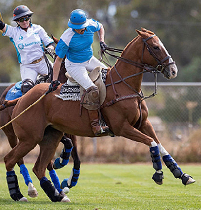
Mare Returns to Polo after Successful Fracture Treatment
Synovial structures such as joints and tendon sheaths are sterile areas protected from the outside environment. If bacteria are introduced into these structures by a wound, very serious infection can occur. Wounds near joints and tendon sheaths should always be treated as an emergency because early diagnosis and appropriate intervention can be life-saving.
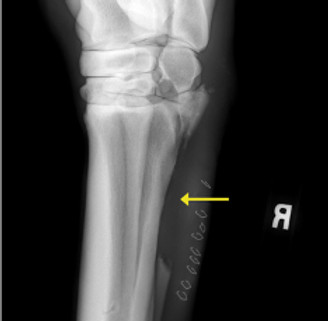
A 9-year-old Thoroughbred Cross mare was presented to one of our Steinbeck Peninsula Equine Clinics veterinarians for a wound just below her hock. Upon evaluation in the field, the horse was grade 4/5 lame in the right hind (lame at the walk). Close examination of the wound along with radiographs (x-rays) showed a comminuted fracture (broken into many small pieces) of the lateral (outside) splint bone with high suspicion of joint involvement.
The mare was promptly transported to the Salinas clinic for further care. Under general anesthesia, wound communication with the tarsometatarsal (lower hock joint) was confirmed and the joint was lavaged (flushed). Loose bone fragments were removed and the laceration was repaired. During surgery, a regional limb perfusion was performed to deliver a high concentration of antibiotics to the affected limb. For this procedure, a tourniquet is placed above the affected region and antibiotics are administered directly into the saphenous vein; the tourniquet is left on for about 25 minutes during which time local antibiotic concentration is very high.
Following the procedure, the horse remained at the clinic for intravenous antibiotic therapy and careful monitoring. She was also treated with non-steroidal anti-inflammatories (NSAIDs), bandaging and rest. The mare did very well and after approximately one week she was transitioned to oral antibiotics and allowed to return home. Thirty days after injury, she began a gradual rehabilitation protocol and five months after injury had fully returned to her previous level of work.
Posted with owner permission.
A sterile cotton swab was used to explore the wound at initial evaluation.

Yellow arrows point to multiple fracture lines on the radiograph from initial evaluation.

The lower hock joint was flushed in surgery.

The yellow arrow on this post-operative radiograph points to where nearly half of the affected splint bone was removed.